

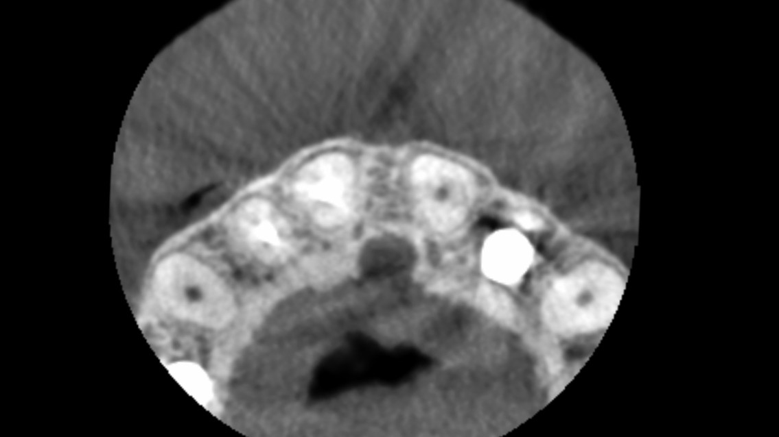
Introduction to Tooth Extraction and Bone Healing
Following tooth extraction, the alveolar bone inevitably undergoes resorption, particularly the buccal plate. This biological process occurs due to the loss of the bundle bone and periodontal ligament (PDL) complex, which is crucial for the vascularization of the buccal plate. Without the tooth in place, the buccal plate resorbs, unless its thickness exceeds 2 mm. However, studies show that a buccal plate thickness of 2 mm is extremely rare—found in only 8.1% of lateral incisors, 2.7% of canines, and virtually never in central incisors.
Conventional Methods and Guided Bone Regeneration (GBR)
Traditionally, to preserve the alveolar buccal bone after extraction, several conventional approaches have been utilized. These include socket preservation, delayed two-stage implant placement, and immediate implant placement accompanied by augmentation. The latter often involves flapless or conventional surgery utilizing bone substitutes and double-layer collagen membranes—a technique widely known as Guided Bone Regeneration (GBR).
While GBR has been a gold standard for decades, it presents significant drawbacks. It requires a complex surgical approach and extensive training. Furthermore, it is associated with a relatively high rate of complications. Literature suggests an average complication rate of around 16.8% to over 25%, depending on the type of membrane used (e.g., e-PTFE, Titanium mesh, or absorbable membranes). Complications such as membrane exposure can lead to bacterial contamination, reduced bone volume, and even partial or complete failure of the bone graft. Additionally, the esthetic outcome with GBR can be unpredictable; achieving a perfect Pink Esthetic Score (PES) is challenging, papillae may be reduced, and the natural appearance of the gingiva can be compromised.
History of the Socket-Shield Technique
In response to the challenges and complications associated with GBR, the Socket-Shield technique was introduced as a part of Partial Extraction Therapy (PET). The technique was first described by Professor Hürzeler in 2010, building upon Professor Salama’s earlier successes with implant-supported bridges over subgingivally retained roots. The rationale is simple yet profound: by retaining a buccal fragment of the root, the periodontium (including the bundle bone and PDL) is preserved, thereby preventing the inevitable resorption of the buccal plate without the need for complex grafting procedures. The term PET and its classification were further formalized by Gluckman and colleagues in 2016.
Procedure Overview
The protocol for the Socket-Shield technique requires meticulous execution. The coronal portion of an unsalvageable tooth is removed, and the root is sectioned to extract the palatal fragment while leaving the buccal “shield” intact. Key guidelines include:
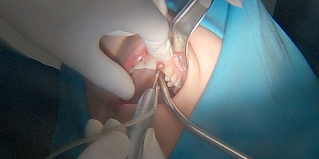
- Shaping the shield to a concave form that naturally follows the preparation.
- Reducing the shield to the bone level, at least 2 mm below the gingival margin.
- Ensuring a thickness of approximately 1.5 to 2 mm.
- Completely removing the root canal to prevent infection.
- Avoiding direct contact between the shield and the implant or prosthetic components to prevent fracture or pressure.
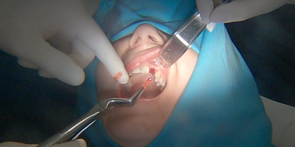
Following preparation, immediate implant placement via a flapless approach is standard, ideally paired with immediate loading if adequate torque (over 35 Ncm) and bicortical anchorage are achieved. The One Abutment One Time protocol and screw-retained restorations are highly recommended to ensure stability and protect the crestal bone.
For a detailed clinical example, you can review our Socket Shield Technique Case.
Supporting Studies and Literature
The efficacy and predictability of the Socket-Shield technique have been supported by numerous clinical studies:
- Hürzeler et al. (2010) provided the proof-of-principle report showing new cementum forming on the prepared root fragment surface.
- Bäumer et al. (2017) reported a 100% success rate with high esthetic outcomes after 5 years, though advising careful execution.
- Gluckman et al. (2018) conducted a pivotal retrospective evaluation of 128 cases over up to 4 years. They reported a high survival rate with minimal issues, categorizing shield exposure into internal and external, with an overall shield exposure rate of 12.5% and a loss of osseointegration at just 3.9%. Gluckman later published a clear step-by-step protocol in 2020 to minimize these complications.
- Systematic reviews, such as those by Xi Lin et al. (2022) and Stefano Oliva et al. (2023), further confirm that the complication rate of the Socket-Shield technique is not statistically higher than conventional immediate placement with GBR, yet the preservation of the buccal bone and esthetics (PES) is significantly better.
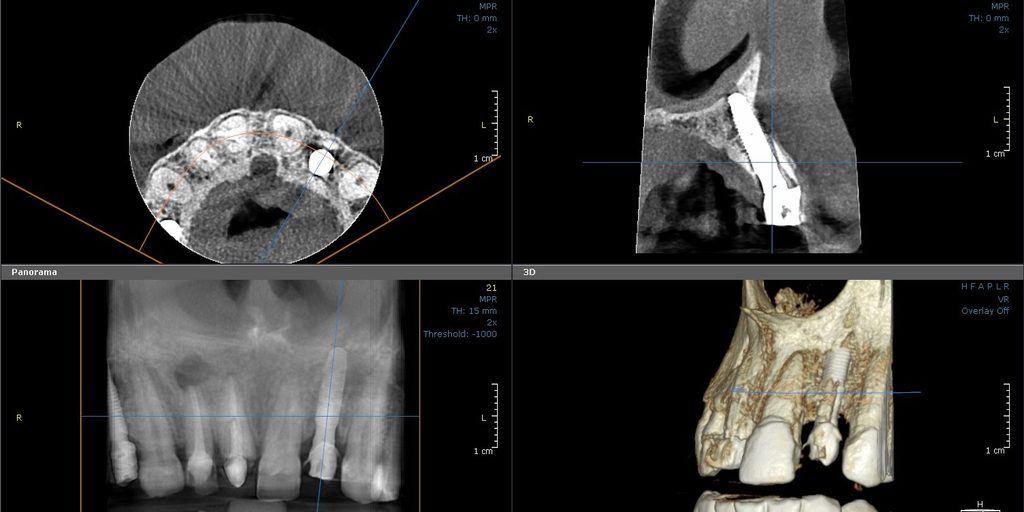
Complications
Despite its advantages, the Socket-Shield technique is not without potential complications. These primarily include:
- Root fragment mobilization.
- Shield exposure: This can be external (gingival) if the fragment is not submerged enough, or internal if there is insufficient space between the fragment and the prosthetic component.
- Root infection.
- Peri-implantitis: Usually secondary to infection of the root fragment.
While most complications (such as shield exposure) can be managed successfully, adherence to strict clinical protocols is crucial to minimize their occurrence.
References
- Hürzeler MB, Zuhr O, Schupbach P, Rebele SF, Emmanouilidis N, Fickl S. The socket-shield technique: a proof-of-principle report.
- Bäumer D, Zuhr O, Rebele S, Hürzeler M. Socket Shield Technique for immediate implant placement – clinical, radiographic and volumetric data after 5 years. Clin Oral Implants Res. 2017;28(11):1450-1458. doi:10.1111/clr.13012
- Gluckman H, Salama M, Du Toit J. A retrospective evaluation of 128 socket-shield cases in the esthetic zone and posterior sites: Partial extraction therapy with up to 4 years follow-up. Clin Implant Dent Relat Res. 2018;20(2):122-129. doi:10.1111/cid.12554
- Gluckman H, Du Toit J, Salama M, Nagy K, Dard M. A decade of the socket-shield technique: a step-by-step partial extraction therapy protocol. Int J Esthet Dent. 2020;15(2):212-225.
- Lin X, Gao Y, Ding X, Zheng X. Socket shield technique: A systemic review and meta-analysis. J Prosthodont Res. 2022;66(2):226-235. doi:10.2186/jpr.JPR_D_20_00262
- Oliva S, Capogreco M, Murmura G, Lupi E, Mariachiara DC, D’Amario M. The socket shield technique and its complications, implant survival rate, and clinical outcomes: a systematic review. J Periodontal Implant Sci. 2023;53(2):99-109. doi:10.5051/jpis.2201780089
- Fontana F, Grossi GB, Fimano M, Maiorana C.Osseointegrated Implants in Vertical Ridge Augmentation With a Nonresorbable Membrane: A Retrospective Study of 75 Implants With 1 to 6 Years of Follow-Up. Int J Periodontics Restorative Dent. 2015;35(1):29-39.
- Lim G, Lin GH, Monje A, Chan HL, Wang HL.Wound Healing Complications Following Guided Bone Regeneration for Ridge Augmentation: A Systematic Review and Meta-Analysis. Int J Oral Maxillofac Implants. 2018;33(1):41-50.
- Urban IA, Montero E, Monje A, Sanz-Sánchez I.Effectiveness of vertical ridge augmentation interventions: A systematic review and meta-analysis. J Clin Periodontol. 2019;46 Suppl 21:319-339.
- Sanz-Sánchez I, Sanz-Martín I, Ortiz-Vigón A, Molina A, Sanz M.Complications in bone-grafting procedures: Classification and management. Periodontol 2000. 2022;88(1):86-102.
- Gaudimier Z, Tawfik S, Kerouanton C, Wulfman C, Flottes Y.Oral bone regeneration and associated complications: A systematic review and meta-analysis. J Stomatol Oral Maxillofac Surg. 2026.


Leave a Reply